Gfap Antibody Cell Signaling

Pin On Antibodies

Gfap D1f4q Xp Rabbit Mab Cell Signaling Technology

Pin On Antibodies

Pin On Cyberpunk

Bioworld Technology Inc Technology Innovative Research Signal Transduction

Pin On Antibodies

We are dedicated to providing innovative research tools that are used to help define mechanisms underlying cell function and disease.
Gfap antibody cell signaling.

Ton10gvd7x3e M

Astrocytes Or Astrocytic Glial Cells Collectively Form Astroglia Star Shaped Cells Surrounding Neurons In The Brain And Sp Glial Cells Neurons Neuropsychology

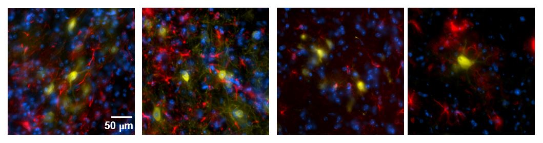
Fluorescent Tagging Is A Powerful Way Of Analyzing Communications Between Cells This Method Enables The Analysis Of Numero In 2020 Confocal Microscopy Cell Microscopy

Gfap Ga5 Mouse Mab 3670s From Cell Signaling Technology Biocompare Com

Pin On Antibodies

Slc2a4 Antibody Has Been Validated For Elisa Wb Ihc If And It Will Be Offered At 9 9 Before December 31 Hurry Biotechnology Muscle Tissue Skeletal Muscle

Confocal Immunofluorescent Analysis Of Rat Brain Using Gfap Ga5 Mouse Mab Alexa Fluor 488 Conjugate Green Textures Patterns Natural Shapes Medical Photos

Anti Gfap Antibody Chicken Anti Rat Glial Fibrillary Acidic Protein Gfap Polyclonal Antibody Np 776490 2

Recombinant Anti Gfap Antibody Epr1034y Ab68428 Abcam

Dna The Ultimate Hard Drive Dna Sequence Biomedical Biomedical Science

Pin By Dan Bose On Science Is Cool Alexa Thermo Fisher Notes

Gfap Antibody Western Elisa Sab2500462 Anti Glial Fibrillary Acidic Protein Sigma Aldrich
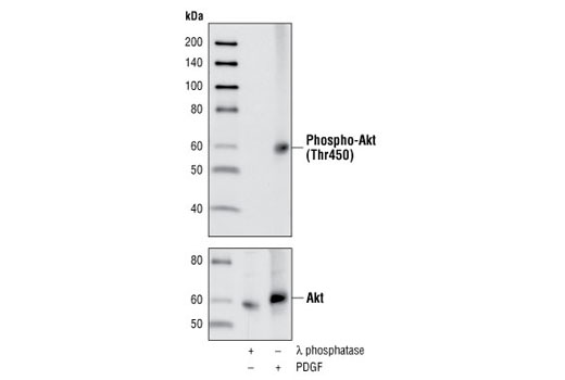
Phospho Akt Thr450 Antibody
Nsc And Astrocyte Marker Antibody Duo Gfap Nestin Arg30006 Arigo Biolaboratories

Goat Anti Rabbit Igg Antibody H L Dylight 488 Heavy And Light Minimal Background Light Chain
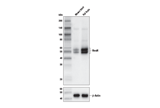
Neun D4g4o Xp Rabbit Mab Biotinylated

Neun D4g4o Xp Rabbit Mab

Asea Science Dr Samuelson Asea Rejuvination Science

Schwann Cells Markers Markers Cell Flow Cytometry
Source : pinterest.com
